Start here · Parmita Mishra
Virtual cells,
explained.
What they are. How they work. Why they matter.
Add a drug. Switch off a gene. Take away oxygen. What will the cell do next?
A virtual cell is a computer model built to predict an answer. The ambition is to test more ideas, understand why they work, and choose better experiments.
The idea is easy to grasp. The hard part is learning enough about a living cell to make a prediction worth trusting.
First, what is a cell?
A cell is the basic unit of life. Inside a tiny boundary, it uses energy, builds and repairs itself, processes information, and responds to its surroundings. It is doing the work of staying alive.
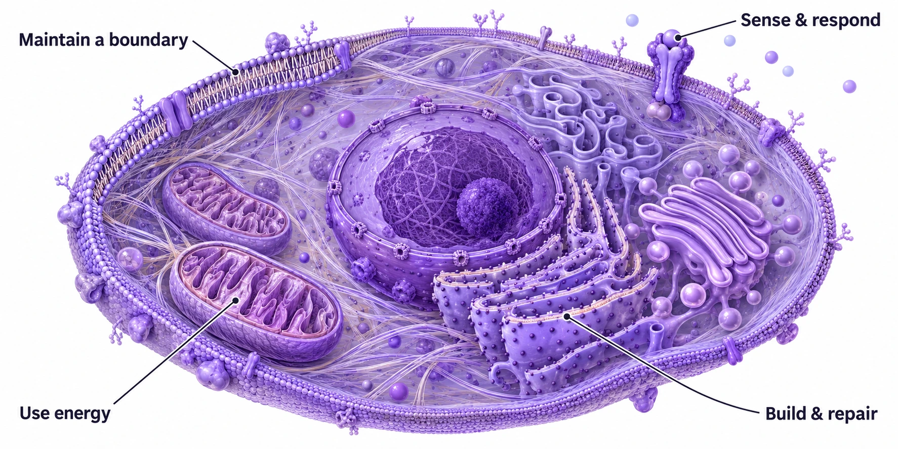
Some cells live as whole organisms. In your body, cells work together as tissues and organs. A liver cell behaves differently from a nerve cell—and the same liver cell can behave differently when it is fed, stressed, or exposed to a drug.
That changing behavior is what we want to understand and predict.
How cells sense, maintain, and repair
A cell senses changes and actively keeps its internal conditions within workable ranges. Biologists call this homeostasis. Michael Levin places this capacity at the beginning of a broader framework for biological intelligence:
Michael Levin · Biophysical Intelligence Between Genotype and Phenotype, 2026
homeostasis is the first rung on the ladder of intelligence
He also writes: Single cells are composite agents that exhibit extremely rich patterning and behaviors
. Read the 2019 paper.
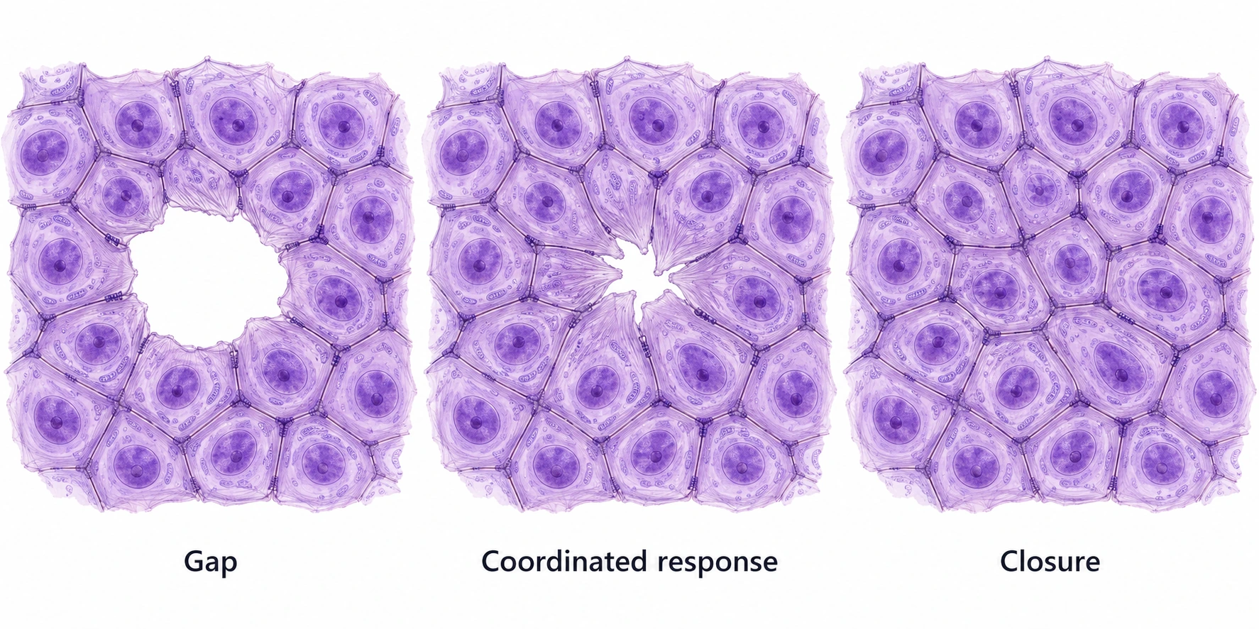
Consider a gap in a sheet of epithelial cells—the cells that line surfaces. Cells at the edge spread and move. Forces and signals coordinate their neighbors. Together, they can close the gap. The behavior belongs to an organized, interacting system.
Liver cells, T cells, and the environments they live in
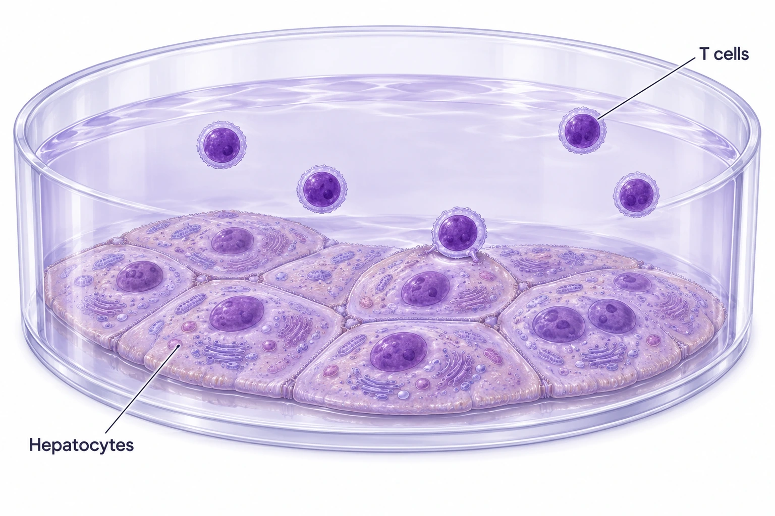
A liver cell, or hepatocyte, processes nutrients and many drugs. A neuron carries signals. A fibroblast helps build connective tissue. A T cell participates in immune responses. “Cell” names a whole world of different jobs.
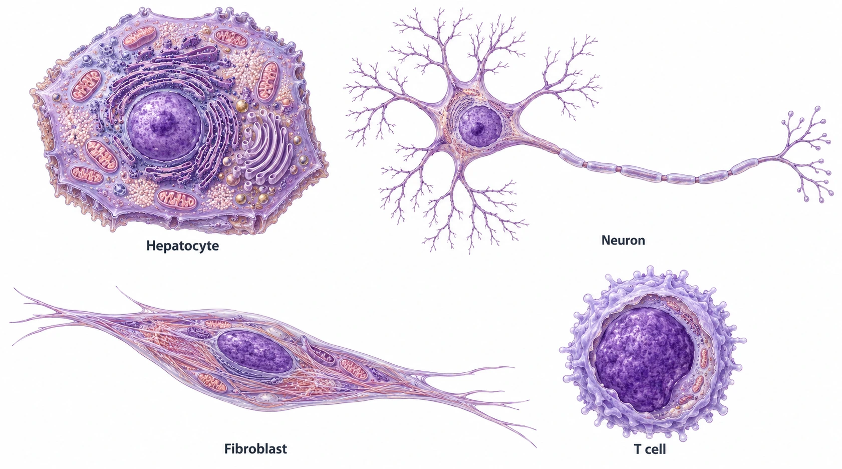
Cell type is its biological identity. Cell state is its condition now. A resting T cell and an activated T cell can share a type while behaving differently.
How we grow cells matters too. In an adherent culture, they attach to a surface. In a suspension culture, they remain in the medium. Plated hepatocytes and suspended T cells are two examples.
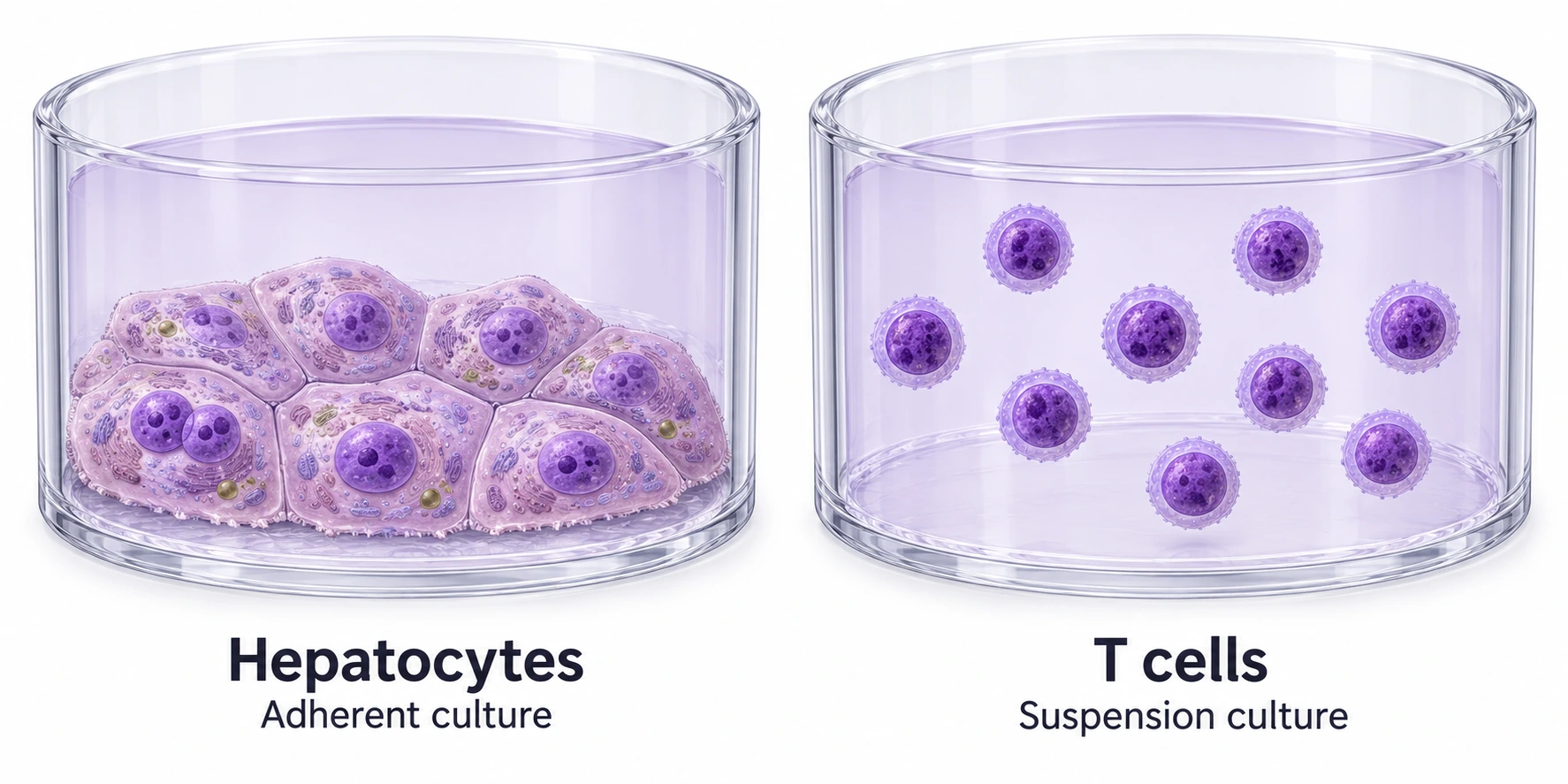
Sometimes we need both. A mixed culture brings tissue cells and immune cells into a shared environment, letting us study how they affect one another.
What is a virtual cell?
A virtual cell is a model you can ask “what if?” You describe a cell and a change. The model predicts a particular response.
Imagine giving a drug to liver cells. You want to know whether they keep functioning, become stressed, or recover after the drug is removed. A model might tackle one part of that question: how RNA—the messages and working molecules copied from DNA—changes after a specified dose and time.
Predicting RNA is useful. Predicting recovery is a further task, with a different test. The phrase “virtual cell” covers models with very different abilities.
The useful question is: what can this model predict, for which cells, under which conditions?
If the prediction is reliable, it can help choose which compound, dose, or experiment to try next. The point is to make experiments more informative.
How does a model learn
what cells do?
For a model that learns from data, the starting point is an experiment: change something, measure what follows, and keep a record of the conditions.
Biologists call the deliberate change a perturbation. It can take several forms.
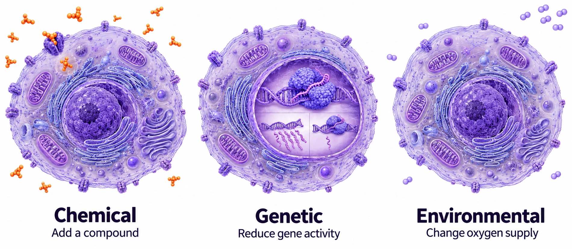
- Chemical
- Add a drug or another compound.
- Genetic
- Change a gene or how active it is.
- Environmental
- Change oxygen, nutrients, temperature, or the surroundings.
First, collect examples. Give cells different treatments and record their responses. The dose, timing, cell type, and comparison condition belong with the data.
Then, train the model. It learns relationships between what changed and what happened. The output could be RNA levels, cell shape, growth, or another measured feature.
Finally, ask a new question. Hold an experiment out of training. Ask the model to predict it, then compare the prediction with the actual result. This is validation.
A model can fit familiar examples and still fail on a new drug or cell type. The test is what it gets right beyond the examples it learned from.
Why is this so hard?
A cell is one connected system. Our instruments give us different views of it.
One method reads DNA. Another counts RNA. Another measures proteins. These large-scale studies are called omics: measurements of whole classes of biological components.
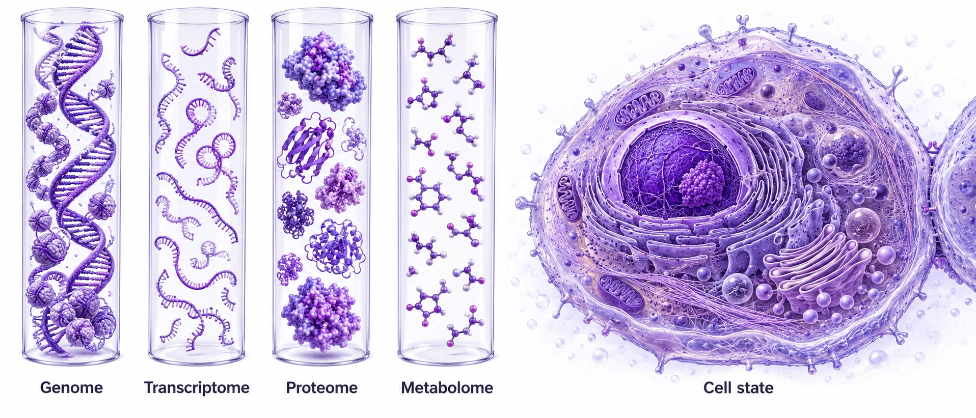
- Genomics
- DNA: genetic information.
- Transcriptomics
- RNA: messages and other working transcripts.
- Proteomics
- Proteins: machinery, signals, and structure.
- Metabolomics
- Small molecules involved in metabolism.
A parts list is not a cell state. The amounts matter, but so do the locations, interactions, surroundings, and activity of those parts.
Think of chess. Knowing which pieces exist does not tell you the position or what happens next. For a cell, we need to understand how its components are organized and working together.
Our data often leave us with a sharded representation—pieces of information about the same living system, collected through different methods and sometimes from different cells.
Multiomics combines several molecular views. Combining molecular measurements with a cell’s shape and behavior gives us a fuller view. The aim is to connect what a cell contains with what it does.
For our liver-cell example, a change in RNA is one clue. To understand recovery, we also need evidence about how the cells function afterward.
The cell has a history.
Return to our drug-treated liver cell. Four hours later, it is alive. Is it recovering? Adapting? Becoming injured? What happens when the drug is removed?
A final reading can miss the route the cell took to get there.
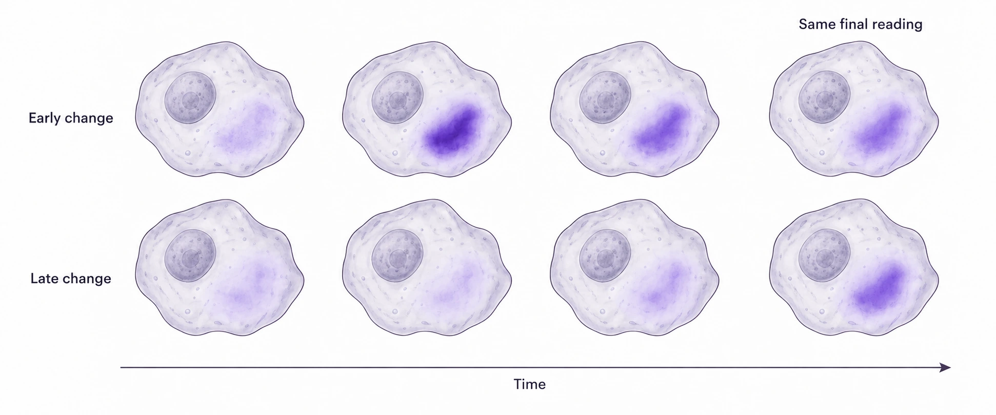
A trajectory is a path of change through time. Did a response begin immediately or after a delay? Did it keep growing? Did it reverse after treatment stopped?
Some models infer these paths from snapshots. Repeated measurements can provide direct evidence about how features change. Following the same individual cell also requires tracking it.
This is where trajectomics comes in.
Trajectomics is the name I use for studying cellular response histories: what changes, where, when, and in what order.
The proposal is to give models richer examples of living responses, then test whether those histories improve prediction. In our drug example, the question is whether the early response helps predict later function or recovery.
Waddington’s landscape: a picture of possible paths
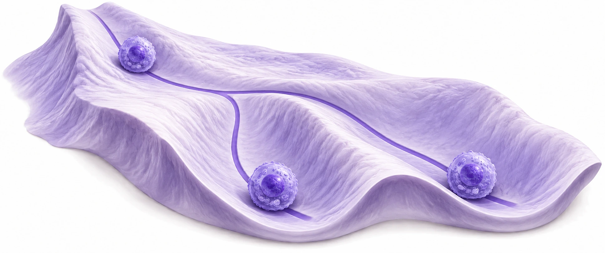
Picture a ball rolling through branching valleys. Its position represents a state; its route represents change; the valleys suggest alternative outcomes. Waddington described the ball’s path as the developmental history of a particular part of the egg
in The Strategy of the Genes (1957, p. 29). His original picture was about development.
Now borrow the metaphor for aging. Imagine following liver cells over time. One path maintains their ability to do their job; another progressively loses function. A hypothetical non-aging reference stays stable. Similar starting points can lead to different histories.
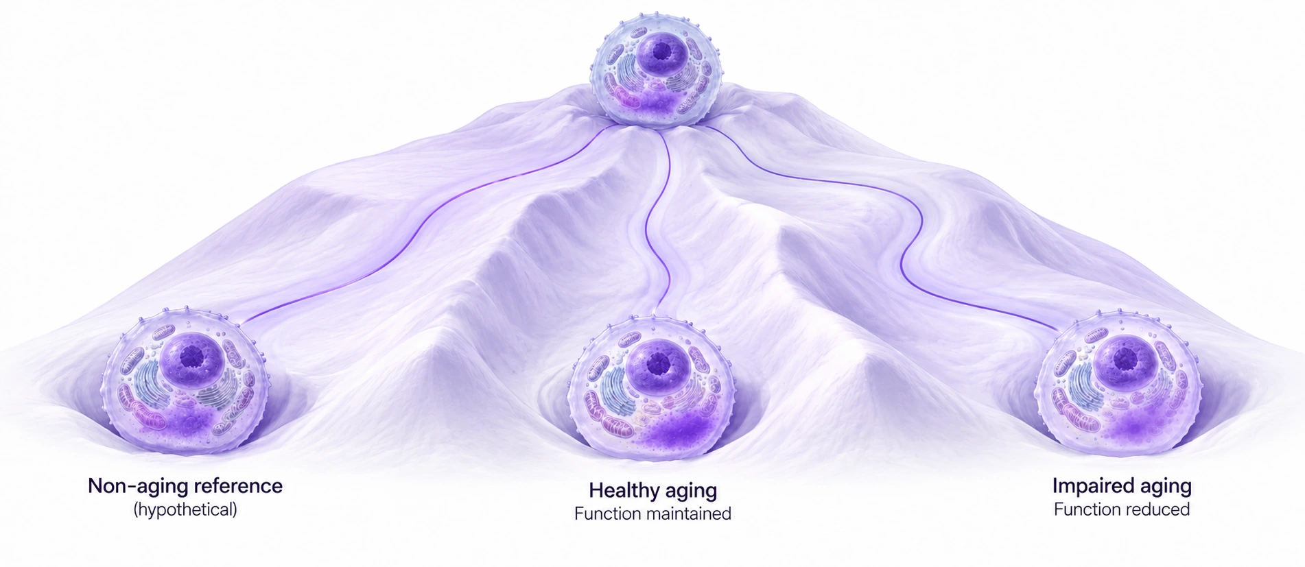
These terrains illustrate an idea. A landscape learned from data is a separate construction whose shape depends on what is measured and how it is modeled.
Watch the response
while it happens.
To learn a response history, we need measurements through time. One approach is to keep cells alive in a controlled environment, introduce a treatment, and observe what changes.
Flowing fresh medium through the chamber—perfusion—lets us control the exposure. Replacing drug-containing medium with drug-free medium is washout. The response may continue after the drug supply stops.
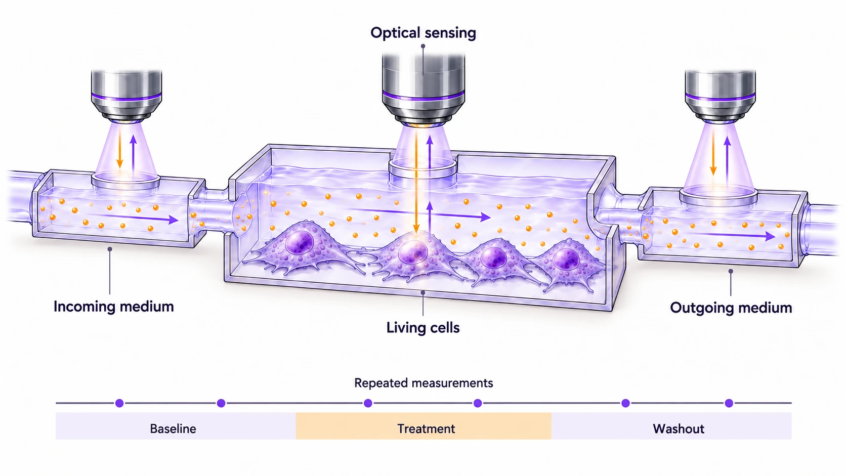
The concept above senses the cells and the medium before and after the chamber. It connects what we deliver with what we observe over time.
This is the measurement direction behind Cell Cinema: living-cell chemical histories that models can learn from. The experiment records the response; a model must still earn its prediction on new data.
How does this compare with destructive sampling?
Conventional destructive single-cell RNA sequencing reads different cells at successive times. A sample leaves the culture vessel. The collected cells are then lysed—broken open—in a separate tube.
Live imaging can return to a retained living field. Both approaches can cover time. One consumes the sampled cells; the other can observe the field again.
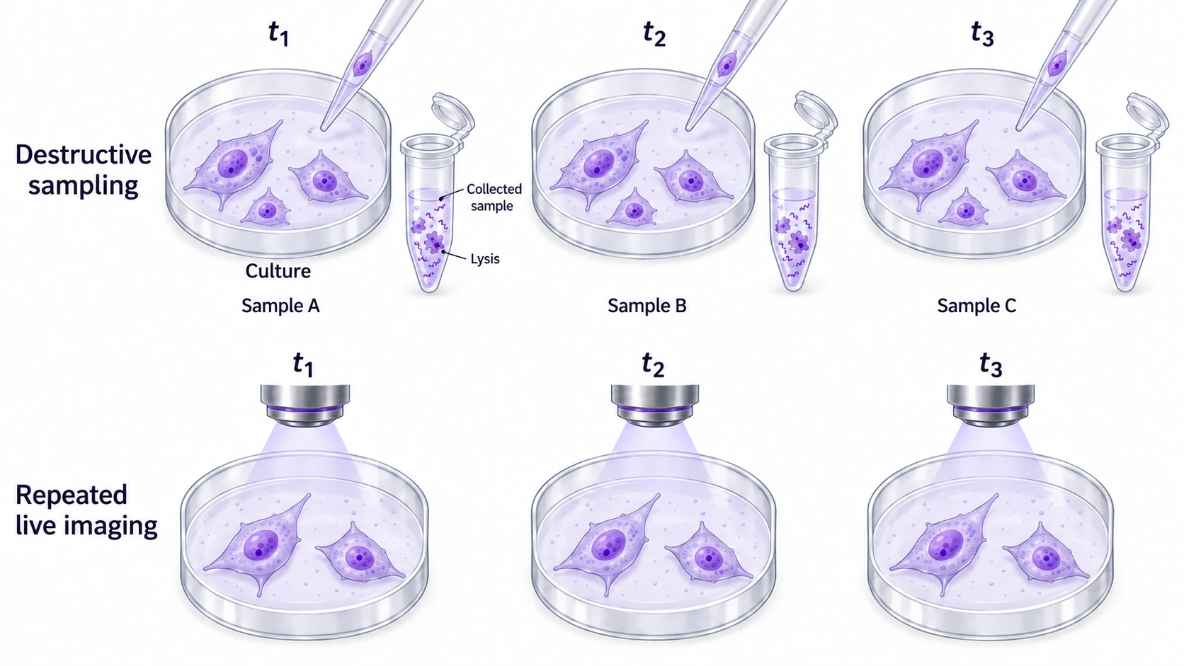
They answer complementary questions and provide different molecular coverage. Specialized methods such as Live-seq can sample RNA while preserving viability. Repeatedly imaging a field is also different from tracking every individual cell within it.
Hyperspectral imaging: chemistry behind each pixel
An ordinary RGB image has three color channels. A hyperspectral image records many spectral channels at each sampled location. Think of an image with a spectrum behind each pixel.
A spectrum records a signal across wavelengths or frequencies. In Raman imaging, its patterns contain chemical information.
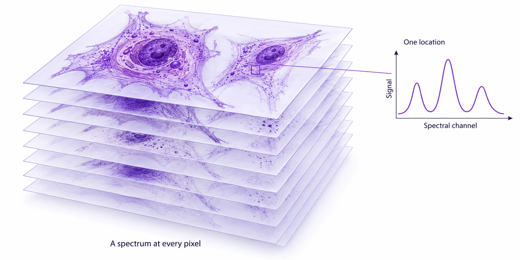
Repeating that measurement can connect chemistry, location, and time. A spectrum is another view of a cell, with overlapping contributions that require interpretation. It is not automatically a complete molecular inventory or a multiomics experiment.
One measured exampleSeven fields in one pilot
In our exploratory “A cell has more than one clock” observations, heme-associated contrast crossed below half its starting value before broad CH contrast did in seven sampled fields. The timing differed across fields.
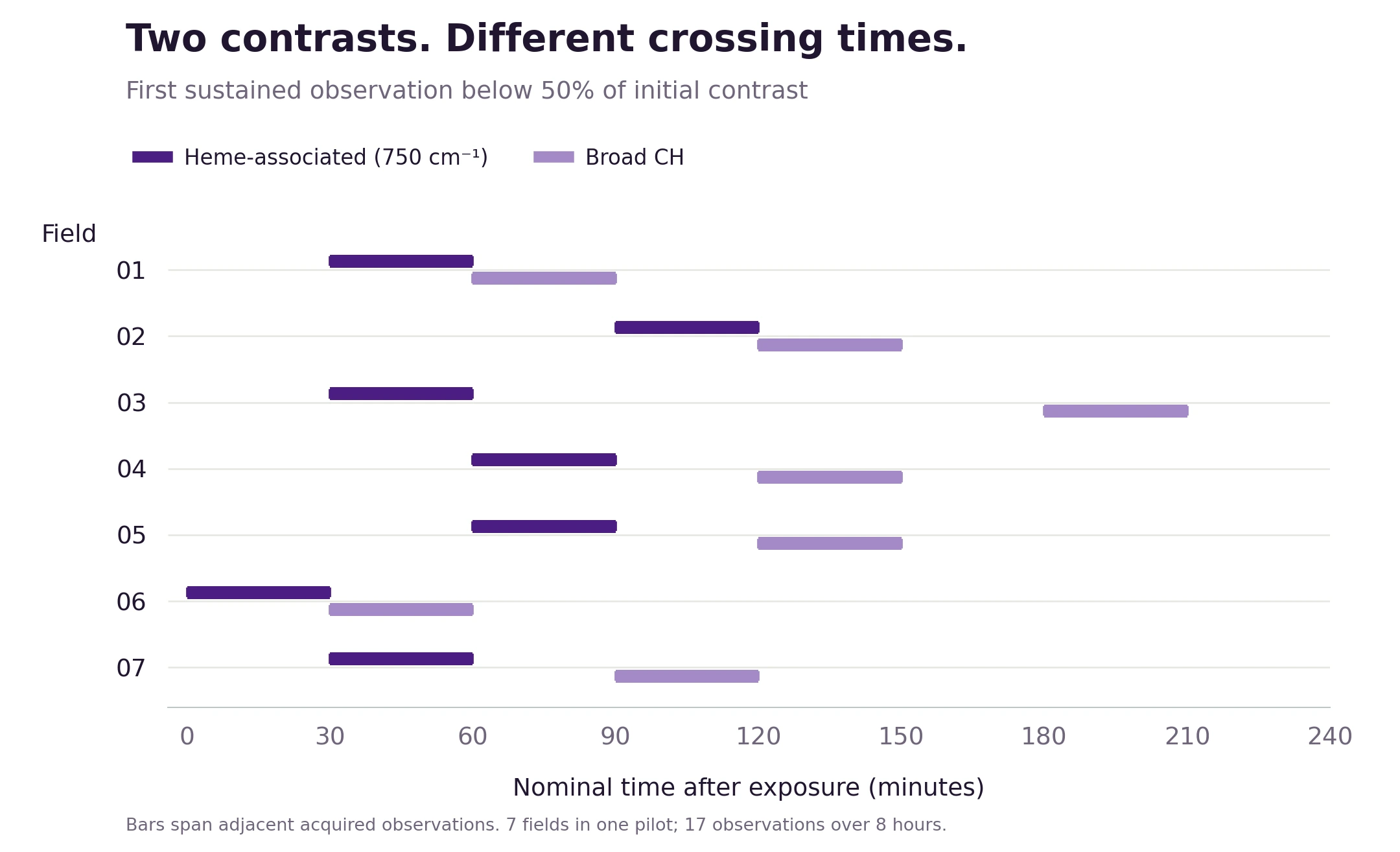
This is an observation about two processed chemical features. Connecting it to injury or recovery requires independent outcomes. Processing choices and the incomplete raw-source flag are in the figure notes.
What can virtual cells
actually do?
They can already answer some well-defined questions. CellBox predicted responses to drug treatments left out of training in a melanoma cell line. PRESCIENT modeled how cells develop and checked predictions about the kinds of cells they become against records linking cells to their descendants.
These are specific successes. The larger challenge is reliable prediction across new treatments, cell types, and environments.
When you hear a virtual-cell claim, ask three things: what did it predict, what was new to the model, and how was the answer checked? Those questions turn a broad promise into something you can evaluate.
And a virtual organ? A virtual human?
Cells communicate and organize into tissues. Tissues form organs. Connecting models across those scales is the larger ambition.
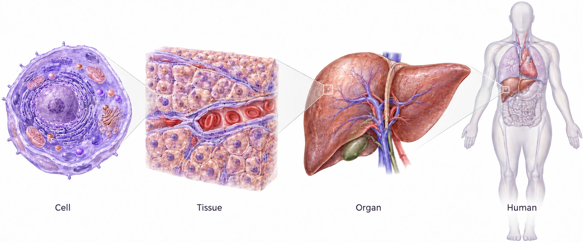
Live-cell observations can help describe how cells change and interact. A virtual liver also needs tissue architecture, transport, and organ function. A virtual human adds communication between organs and the rest of the body.
The AI virtual-cell perspective sets out this direction. The path forward is to build capabilities that can be tested, then connect them.
Keep exploring
MotivationHMCVelo & the piano
My HMCVelo work uses joint measurements of DNA methylation and hydroxymethylation to infer epigenetic velocity from single-cell snapshots. It motivates a related question: what can be inferred from a snapshot, and what more can be learned by observing change?
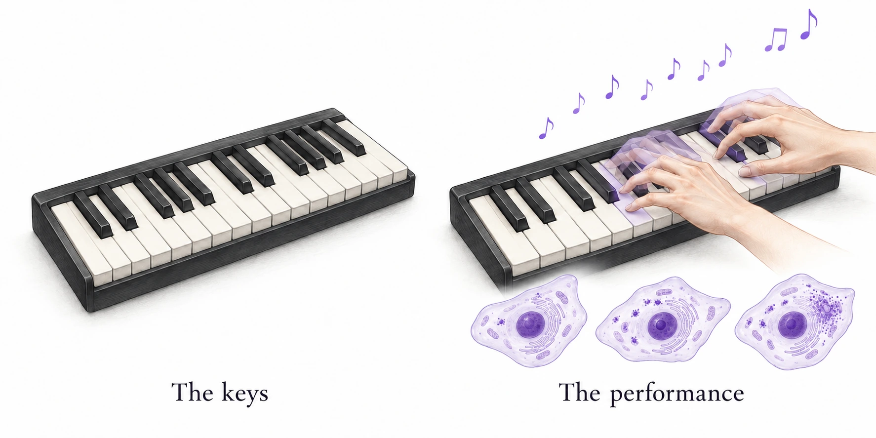
My piano thread offers an analogy. Genes are like keys. Which ones are played, together, and when? The analogy is a starting point for thinking about coordinated activity over time.

The idea to take with you
Predict what a cell will do.
Then ask the living cell.
Better models need better experiments.
Better experiments can teach us what to model.